What is Cancer?
Cancer is a class of diseases in which a group of cells display
- uncontrolled growth (division beyond the normal limits),
- invasion (intrusion on and destruction of adjacent tissues) and
- metastasis sometimes (spread to other locations in the body via lymph or blood).
These three malignant properties of cancers differentiate them from benign tumors, which are self-limited lesions that do not invade or metastasize.
Cancers often also have a fourth property: angiogenesis, the ability to stimulate new blood vessel growth from pre-existing vessels. This allows cancer cells to obtain their necessary oxygen and nutrients from these blood vessels even when individual members are too sparsely distributed to be connected by diffusion alone.
Because angiogenesis is central to cancer growth and metastasis, it has become a target for the treatment of cancer.
What are the Most Common Types of Cancer?
The most common types of cancer are: prostate, breast, lung and bronchus, colon and rectum, bladder, non-Hodgkin lymphoma (NHL), pancreas, melanoma of the skin, kidney and ureter.
Only about 1% of cancers occur in children
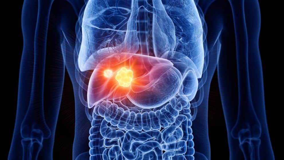
What is the Liver?
The liver is one of our vital organs. It has a wide range of functions, including detoxification, protein synthesis, and production of biochemicals necessary for digestion. The liver is responsible for about 500 separate functions. One of its important tasks is to remove various toxins from the body. This occurs via two mechanisms: secretion into bile or metabolism by liver cells (hepatocytes). All these processes are closely interconnected and contribute to homeostasis. The liver also produces insulin-like growth factor 1 (IGF-1), which stimulates cell proliferation and inhibits apoptosis (cell death) in many tissues such as muscle, bone marrow, kidney epithelium and nerve cells, thus allowing uncontrolled tissue growth
What is Liver Cancer?
The term “cancer” usually refers to malignant neoplasms showing uncontrolled cell division in parts of the body far from their origin. These are called primary cancers or malignant tumors. In contrast, non-neoplastic diseases involving defects in normal cell division such as benign tumors (which do not invade nearby tissues nor spread to distant sites) represent a different category of disease that does not fall under this definition of cancer.
About 90% of all cancers come from epithelial cells: cells that have relatively little mobility and form flat sheets which cover surfaces or line cavities. Such kinds of cells occur in the skin, breast, stomach, oesophagus and other organs. Other common cancers arise from mesenchymal cells: cells with more extensive mobility which give rise to fibrous tissue, muscle and bone. These include leukaemias (cancers of the blood-forming tissues such as the lymphocytes), multiple myeloma (cancer of certain immune system cells) and sarcomas (cancers of connective tissue such as bone or cartilage).
What is Primary Liver Cancer?
Liver cancer can be a primary cancer originating in the liver (for example hepatocellular carcinoma or cholangiocarcinoma) or a secondary tumor metastasizing there from another site in the body. Thus, it is not always easy to know whether a new tumor in the liver is a metastasis or a “true” primary malignancy unless a biopsy is performed.
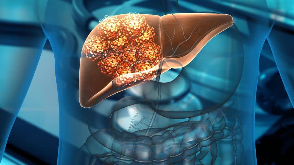
Nearly all cases of primary liver cancer are hepatocellular carcinomas (HCC). Many other kinds of carcinoma, sarcoma and even some leukemia and lymphoma belong to the group of cancers arising from hepatocytes.
Some degree of confusion may arise when a tumor that arose elsewhere metastasizes to the liver or when a primary malignancy originating in the liver (hepatoma) spreads to distant organs such as lungs or bones. The pathologist must make an effort to discover if such tumors began elsewhere before invading the liver or if they actually originated there before spreading elsewhere.
Primary liver cancer (cancer that starts in the liver) is not common. It usually happens because of long-term liver disease (such as cirrhosis). The main types are hepatocellular carcinoma, cholangiocarcinoma, and angiosarcoma.
What is Hepatocellular Carcinoma?
Hepatocellular carcinoma is a common form of primary liver cancer that begins with a tumor in a single cell in the liver. Hepatic means related to or affecting the liver. A hepatocyte refers to a mature liver cell. Carcinomas most commonly begin as growths or swellings called tumors that have formed from cells that have been damaged.
What are Risk Factors for Hepatocellular Carcinoma?
Risk factors for hepatocellular carcinoma include:
* age (greater than 45 years of age)
* male sex
* hepatitis B infection (most common risk factor in Asia and Africa)
* hepatitis C infection (most common risk factor in Europe and North America)
- alcoholic liver disease – related cirrhosis;
- non-alcoholic steatohepatitis
- other: drug toxicity; anorexia nervosa; hemochromatosis; alpha-1-antitrypsin deficiency; Wilson disease, atohepatitis or obesity.
What is Cholangiocarcinoma?
Cholangiocarcinoma is a cancer of the bile ducts. Bile flows through a series of tubes called a biliary tree, where some chemicals and waste products in the bloodstream are changed as they go through liver, pancreas and intestine. This helps make them less harmful before they leave your body as waste product in stool. Cholangiocarcinoma often starts in cells that line the inside bile ducts. This process may have been started by chronic inflammation.
What is Liver Angiosarcoma?
Liver angiosarcoma is rare type of cancer that arises from the blood vessels inside the liver. Liver contains multiple large blood vessels through which it receives nutrients and oxygen, as well as disposes of toxins. Normally these blood vessels are durable, but in some cases they can be fragile, leading to leakage which fills space between liver cells with liquid or air. This condition is known as hemoperitoneum. If this occurs for prolonged periods, it leads to development of tumour-like mass made up of abnormal tissues similar to those found in skin or other organs.
How Does a Liver Cell Become Cancerous?
Cancerous changes result from mutations – permanent changes in the DNA of a cell. DNA carries a person’s entire genetic information and determines a cell’s characteristics, including whether it becomes a liver cancer cell or a healthy liver cell. Mutations can be inherited from parents, occur during a person’s lifetime due to exposure to harmful substances such as tobacco smoke or ultraviolet rays from sunshine, or result from errors that happen when cells divide in mitosis or meiosis (cellular processes involved in reproduction).
How does Cirrhosis Cause Liver Cancer?
Cirrhosis, or chronic liver disease is the result of long-term damage to liver cells. Often it has no known cause, but heredity may play some role. Alcohol abuse is the most common cause of cirrhosis in Western countries. Hepatitis infection can also lead to cirrhosis. The harmful effect on liver cells results from a combination of factors including loss of certain blood constituents (due to blockage), decreased oxygen supply and inflammation.
Cirrhosis causes hepatocellular cancer by leading to repeated cycles of liver cell damage and regeneration, which may occasionally introduce mutations that initiate tumour growth. Loss of cells in cirrhosis means there is less liver tissue available to perform vital functions. If loss becomes extensive, it results in potentially life-threatening conditions like bleeding from dilated veins or fluid accumulation in the abdomen (ascites).
What are the Symptoms of Primary Liver Cancer?
The most common early symptom is painless swelling of the abdomen. Other signs may include yellowing of the skin and eyes (jaundice), easy bruising bleeding, fatigue, loss of appetite and weight, fever and chills.
How is Liver Cancer Diagnosed?
Liver cancers can be difficult to detect early because there may not be any symptoms for a long time. However, people with risk factors such as cirrhosis or chronic viral hepatitis should get regular blood tests to look for evidence of liver damage. Ultrasound scans (sonography) can help to spot tumours in the liver which produce specific echoes that indicate their presence. Liver function tests are also useful but high concentrations of bilirubin can also cause similar scan readings so they have to be interpreted with caution. If needed, a biopsy can determine if abnormal cells are cancerous by taking tissue samples from the liver using fine needles inserted through the skin on the tumour mass. This is most commonly performed by radiological guidance (using X-rays).
What is Secondary Liver Cancer?
Secondary (metastatic) liver cancer is when the disease spreads to the liver from another location in the body. The original cancer can spread in many ways: through blood or lymph vessels, by direct extension of the original tumor, or metastasizing outside the circulatory system. A common site for such metastases is in nearby lymph nodes and sometimes in bone marrow.
What are Risk Factors for Secondary Liver Cancer?
Risk factors for secondary cancers include:
- primary lung cancer
- breast cancer
- colon and rectal cancers
- stomach and esophageal cancers and others may spread to bones and/or other organs before they reach the liver
How Common are Liver Metastases?
Liver metastases are particularly common in patients with lung cancer, but may also occur with cancers of the breast, colon and rectum. The prevalence of seondary liver metastases is estimated at 30-40 percent of patients with metastatic lung cancer.
How is a Liver Cancer Diagnosis Made?
A diagnosis of liver cancer is usually made by tests (eg biopsies) to determine whether there are any abnormal cells in the liver. The doctor will ask about your medical history, including risk factors such as previous lung or other cancers. The doctor will also do a physical examination and may order certain blood tests. Imaging tests that may be performed include ultrasound, computed tomography (CT) scans, magnetic resonance imaging (MRI), positron emission tomography (PET) scans and angiograms.
How can an MRI Tell You If You Have Hepatocellular Carcinoma (Primary Liver Cancer) Using the Li-Rads Criteria?
An MRI with contrast material is performed using LI-RADS criteria. Sometimes the doctor may inject contrast material into your veins to make the liver tumors easier to see on an MRI.
What Are LI-RADS?
LI-RADS stands for Liver Imaging Reporting and Data System which are guidelines for reporting abnormal findings on MRIs of the liver. These are rules that have been determined based upon consensus by multiple medical groups, to ensure that MRIs get read uniformly across radiologists so they can be read accurately, quickly and with less confusion.
A LI-RADS 5 lesion on MRI means you have hepatocelluar cancer. Less than a score 5 may require a biopsy.
(non-surgical alternatives) advanced liver cancer treatments
How is Liver Cancer Treated?
Treatment for liver cancers depends on the type of cell involved, whether it has spread to other tissues, its degree of malignancy or grade, how large it is and whether there are any symptoms. Treatment may involve:
- surgery in a small number of cases where the tomor can be resected
- chemo- or immuno- therapy from a medical oncologist
- advanced targetted treatment from an interventional radiologist using miniatuized equipment to treat tumors directly
- radiotherapy in some cases provided by a radiation oncologist
A multidisciplinary team approach is usually recommended with the team comprising:
- surgical oncologist
- medical oncologist
- intervention radiologist
- radiation oncologist
- gastroenterologist, pathologis

What are Advanced Liver Cancer Treatment Methods?
Imaging and Interventional Specialists provide advanced liver cancer treatments using state-of-the-art techniques designing to maximally impact liver cancers.
How Do Imaging & Interventional Specialists Determine the Best, Integrated Long-Term Treatment Plan For Your Liver Cancer?
The Imaging & Interventional Specialists team are nationally recognized experts in liver cancer treatment. In fact, Imaging & Interventional Specialists physicians were the first to perform advanced liver cancer (Y90-treatment) in El Paso and are the only providers to use the most advanced Y90 treatment. Besides a careful history and physical exam, the team at Imaging & Interventional Specialists will obtain imaging and lab results and communicate with other members of your treatment team to put together the best, integrated, long-term treatment plan.
What Advanced “Pinhole” Procedures Does Imaging & Interventional Specialists Offer for Liver Cancer?
Imaging & Interventional Specialists offer “pinhole” procedures to address and cure pelvic congestion syndrome.
- Transarterial embolization: Your arteries are used like roads to guide specialized equipment next to the tumor in yor liver. Then tiny beads are injected blocking the tiny arteries that feed the tumor This “starves” the liver tumor and so it dies.
- Transarterial chemoembolization: Again, your arteries are used like roads to guide specialized equipment next to the tumor then beads with a powerful tumor-killing agent are injected blocking the tiny arteries that feed the tumor and delivering the agent. This “starves” and “poisons” the tumor and so it dies.
- Transarterial radioembolization: Again your arteries are used like roads to guide specialized equipment next to the tumor then beads with a powerful radioactive agent are injected blocking the tiny arteries that feed the tumor and delivering the agent. This “starves” and “irradiates” the tumor and so it dies.
- Thermal ablation: An needle is placed directly into your liver tumor and either cold or heat is produced at the end of the needle which kills the tumor.
These methods are often done in sequence over a period of time so a long-term treatment is provided.
Imaging & Interventional Specialists “Pinhole” Procedures: Fast Recovery, Less Risk, Less Pain, Comfortable Setting
Our experienced board-certified specialists successfully perform “pinhole” procedures every day offering this region leading, state-of-the-art solutions to pelvic congestion syndrome. Our non-surgical image guided procedures are cutting edge technology without the cutting, without a scalpel.
- Procedures are usually done in a comfortable outpatient setting with familiar friendly staff
- Pinhole procedures offer fast recovery, less risk, and less pain

Why Imaging & Interventional Specialists?
Imaging & Interventional Specialists are leaders in interventional radiology and experts in non-surgical advanced liver treatments
Using worldclass, state-of-the-art equipment, our experienced board-certified specialists are focused on your best outcome.